Head CT Scan Procedure in Post-Traffic Accident Head Trauma Cases: A Case Study at Siloam Sriwijaya Hospital, Palembang
DOI:
https://doi.org/10.52120/jlik.v4i1.107Keywords:
Head CT-Scan, Head Trauma, Non-Contrast CT, Radiology ProtocolAbstract
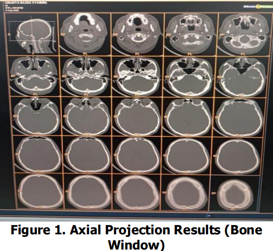
Head trauma resulting from traffic accidents is an emergency condition that requires rapid imaging to exclude life-threatening intracranial injuries. This study aims to describe the procedure and findings of non-contrast head computed tomography (NCCT) in a post-traffic accident head trauma case at the Radiology Department of Siloam Sriwijaya Hospital, Palembang. A descriptive case study design was applied. Data were obtained from NCCT images and validated radiology reports of a 25-year-old male patient examined on May, 2025, complemented by structured interviews with radiographers regarding technical protocols and workflows. The examination was performed using 120 kV, 350 mAs, 5-mm slice thickness for axial acquisition with 1–2 mm multiplanar reconstruction, a field of view of approximately 250 mm, a 512 × 512 matrix, and brain (WL/WW ≈ 40/80 HU) and bone window (WL/WW ≈ 400/2000 HU) settings. NCCT findings demonstrated no intracranial hemorrhage, midline shift, or mass effect. The main pathological findings were nasal bone fracture, septal deviation, and hemosinus involving the bilateral ethmoid sinuses and the left maxillary sine. The applied protocol was consistent with emergency radiology practice and adhered to the ALARA principle through appropriate parameter optimization and multiplanar reconstruction. These findings highlights the role of NCCT US a reliable first-line modality for rapid exclusion of critical intracranial injury while accurately detecting facial bone trauma in traffic accident-related head injuries.
Downloads
References
Ainsworth, C. R., & Brown, G. S. (2021). Head Trauma Guidelines. Journal of the American College of Radiology.
BPS, (2025). Statistik Jumlah Kecelakaan, Korban Mati, Luka Berat, Luka Ringan dan Kerugian Materi.
Coelho, S., Dinis, M. D. L., & Freitas, M. (2026). Radiation Dose Reduction in CT Exams with Iterative and Deep Learning Reconstruction : A Systematic Review. Applied Sciences, 1–26. https://doi.org/10.17605/OSF.IO/TUQDS
Flammia, F., Chiti, G., Trinci, M., Danti, G., Cozzi, D., & Grassi, R. (2022). Optimization of CT protocol in polytrauma patients : an update. European Review for Medical and Pharmacological Sciences, 2543–2555.
Khan, M. S., Alam, M. S., Ismail, S., Ghafoor, B., Sajjad, N., Khan, N., Memon, W. A., Ameen, A. M., Khan, F., Mumtaz, H., Iqbal, J., & Ashraf, A. (2023). Use of National Institute for Health and Care Excellence head injury guidelines among patients with delayed presentation after head trauma can lead to missed traumatic brain injury: a 5-year institutional review. Annals of Medicine and Surgery, 85(9).
National Institute for Health and Care Excellence (NICE). (2023). Head injury : assessment and early management.
Permana, P., Bagus, A., Satyarsa, S., Putu, D., & Wardhana, W. (2025). Karakteristik klinis cedera otak traumatik sedang dan berat di Rumah Sakit Umum Bangli: Studi retrospektif tahun 2024. Medicina, 56(2), 52–57.
Pollock, J. M., Prall, J. A., Ptak, T., Raksin, P. B., Shaines, M. D., Tsiouris, A. J., Utukuri, P. S., Wang, L. L., & Corey, A. S. (2021). ACR
Prabsattroo, T., Wachirasirikul, K., Tansangworn, P., & Punikhom, P. (2023). The Dose Optimization and Evaluation of Image Quality in the Adult Brain Protocols of Multi-Slice Computed Tomography : A Phantom Study. Journal of Imaging.
Pula, Michal, Kucharczyk, Emilia, Zdanowicz- Ratajczyk, Agata, Dorochowicz, Mateusz, & Guzinski, Maciej. (2025). Deep learning and iterative image reconstruction for head CT: Impact on image quality and radiation dose reduction—Comparative study. The Neuroradiology Journal
Putri, P. H., Putri, I. U., Agiestya, M. M., Noor, D., Paranggai, E. A., & Windiyani, F. (2024). Cedera Kepala Sedang setelah Kecelakaan Lalu Lintas (KLL) Tunggal : Sebuah Laporan Kasus. Medula, 14, 518–522.
Qasrawi, R., Thwib, S., Issa, G., Qdaih, I., Ghoush, R. A., & Arjah, H. (2025). Optimized Hounsfield Units Transformation for Explainable Temporal Stage-Specific Ischemic Stroke Classification in CT Imaging. Journal of Imaging, 1–19.
Shih, R. Y., Burns, J., Ajam, A. A., Broder, J. S., Chakraborty, S., Kendi, A. T., Lacy, M. E., Ledbetter, L. N., Lee, R. K., Liebeskind, D. S., Appropriateness Criteria® Head Trauma: 2021 Update. Journal of the American College of Radiology : JACR, 18(5S), S13–S36.
Singh, S., & Sukkala, R. (2021). Evaluation and comparison of performance of low-dose 128- slice CT scanner with different mAs values: A phantom study. Journal of Carcinogenesis, 20, 13.
Tsiouris, A. J., & Lui., Y. W. (2024). Neuroimaging Update on Traumatic Brain Injury. In Diseases of the Brain, Head and Neck, Spine 2024-2027: Diagnostic Imaging. StatPearls Publishing.
Willacy, H. (2024). CT head scanning indications. Patient.Info, 1–8.
Downloads
Published
Issue
Section
License
Copyright (c) 2026 M.Hasbiansyah Putra Wibisono (Author)

This work is licensed under a Creative Commons Attribution-ShareAlike 4.0 International License.